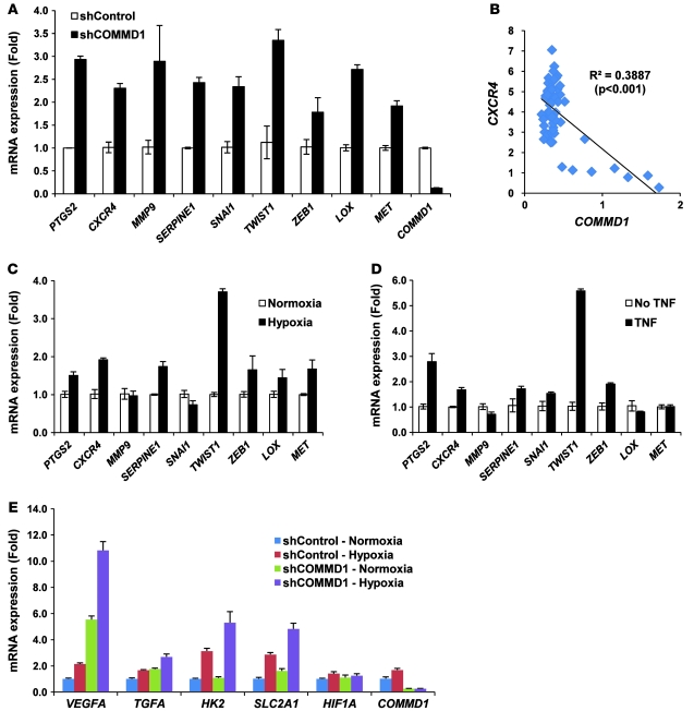
Figure 3. COMMD1 inhibits the expression of HIF-regulated genes.
(A) Gene expression for a panel of invasion-promoting genes was determined in HT29 cells by quantitative RT-PCR and expressed as fold over the shControl sample (mean ± SEM of triplicate samples are shown). (B) Normalized COMMD1 and CXCR4 expression in breast stromal cells (shown in Figure 1E) was plotted and a Pearson’s correlation value was calculated. (C and D) The expression of the same genes was examined in HT29 cells exposed to (C) hypoxia or to (D) the NF-κB activator, TNF. The respective mRNA levels were determined by quantitative RT-PCR and expressed as fold over the untreated sample (mean ± SEM of triplicate samples are shown). (E) Expression of selected HIF target genes was determined by quantitative RT-PCR analysis and expressed as fold over the shControl sample (mean ± SEM of triplicate samples are shown).

