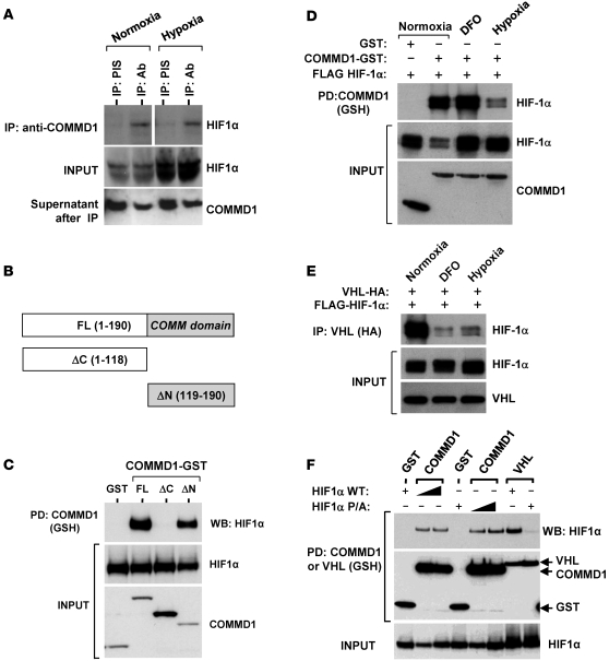
Figure 5. COMMD1 binds to HIF-1α preferentially under normoxic conditions.
(A) Endogenous COMMD1 immunoprecipitation using HEK 293T whole cell lysates incubated either under normoxia or hypoxia (for 1 hour). Whole cell lysates and immunoprecipitates were subjected to Western blot analysis. Supernatants after IP demonstrate immunodepletion of COMMD1. The lanes on the top panel were run on the same gel but were noncontiguous. (B) Truncation mutants of COMMD1. FL, full-length; ΔC, deletion of the carboxyl terminal COMM domain; ΔN, deletion of the amino terminus. (C) These proteins were coexpressed with HA-tagged HIF-1α in HEK293 cells and subsequently precipitated from cell lysates. Coprecipitation of HIF-1α with COMMD1 full-length and ΔN is shown. (D) HEK 293T cells expressing COMMD1-GST, GST, or FLAG–HIF-1α were cultured under normoxia (20% O2) or hypoxia (1% O2, 8 hours) or treated with DFO (0.1 mM, 8 hours), prior to glutahione-sepharose (GSH) precipitations and Western blot analysis as indicated. (E) HEK 293T cells expressing HA-VHL and FLAG–HIF-1α were cultured under normoxia, hypoxia, or treated with DFO as before. Immunoprecipitation of VHL (using anti-HA antibody) was followed by Western blot analysis. (F) HEK 293T cells were transfected with GST, COMMD1-GST, or GST-VHL, along with wild-type HIF-1α or HIF-1α P/A. Subsequently, GSH precipitations were followed by Western blot (WB) analysis as indicated. PD, pull down; PIS, preimmune serum.

