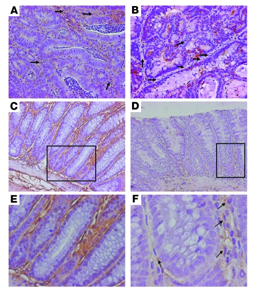
Figure 10. IL-6 expression in Epim–/– and WT colon after treatment with AOM/DSS.
Sections of colon were incubated with goat anti–IL-6 antibody (1:50 dilution). Representative sections of dysplastic (A and B) and nondysplastic (C–F) WT (A, C, E) and Epim–/– (B, D, F) colons are shown. (A) WT dysplastic colon showed light brown stromal staining (arrows), and scattered epithelial cells were positive for IL-6. (B) Dysplastic Epim–/– colon exhibited less stromal staining (arrows), and scattered brown epithelial cells were seen. (C and D) Nondysplastic Epim–/– colon (D) demonstrated less stromal IL-6 than did WT (C). (E and F) Higher-magnification views of boxed regions in C and D, showing intense cellular and extracellular stromal staining in nondysplastic WT colon (E) and markedly reduced staining in Epim–/– colon (F). Scattered pericryptal cells in Epim–/– colon showed persistently positive staining (arrows). Original magnification, ×200 (A–D); ×400 (E); ×1,000 (F).

