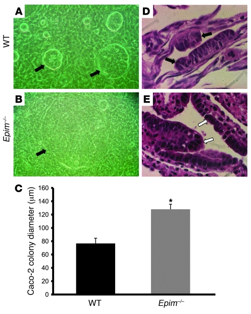
Figure 4. Epimorphin deletion in colonic myofibroblasts alters the morphology of cocultured epithelium.
(A and B) Caco-2 cells were seeded on top of WT (A) and Epim–/– (B) colon myofibroblasts. Growth and morphology were examined daily with an inverted microscope and photographed on coculture day 4. Caco-2 cells (arrows) cultured on WT myofibroblasts formed small, compact colonies with a central hyperdense area; in contrast, Caco-2 cell colonies cultured on Epim–/– myofibroblasts were larger. Original magnification, ×1,000. (C) Mean diameter of Caco-2 colonies grown on Epim–/– compared with WT myofibroblasts: Epim–/–, 128 μm; WT, 76 μm. (D and E) Histologic analysis of H&E-stained myofibroblast–Caco-2 cocultures. Caco-2 cells cocultured with WT myofibroblasts formed predominantly spherical, compact, multilayered, pseudostratified epithelial colonies (D, black arrows indicate epithelium). In contrast, Caco-2 cells cocultured with Epim–/– myofibroblasts formed an epithelial monolayer (E, white arrows). Original magnification, ×1,000. *P < 0.05.

