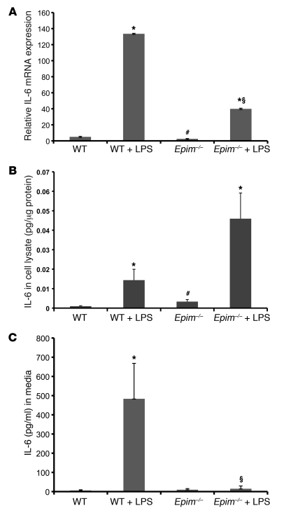
Figure 9. LPS-stimulated IL-6 secretion is altered in Epim–/– myofibroblasts.
(A) IL-6 mRNA expression in WT and Epim–/– myofibroblasts, measured 24 hours after incubation with LPS (5 μg/ml). (B) IL-6 protein expression was stimulated by LPS. IL-6 protein expression in WT and Epim–/– myofibroblasts was measured by ELISA after a 24-hour incubation with LPS (5 μg/ml). IL-6 expression was higher in LPS-treated Epim–/– myofibroblasts than in LPS-treated WT myofibroblasts, although the difference was not significant (P = 0.07). (C) IL-6 protein secretion was markedly reduced in LPS-treated Epim–/– myofibroblasts. Cells were incubated with LPS (5 μg/ml) for 24 hours. *P < 0.05 versus respective untreated group; #P < 0.05 versus untreated WT; §P < 0.05 versus LPS-treated WT.

