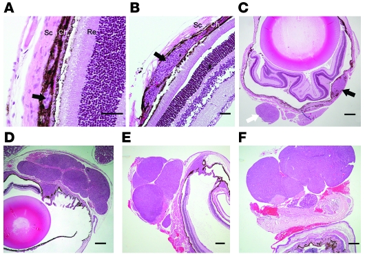
Figure 6. Morphology of eye tumors at various ages.
Eye sections from 2- (A), 4- (B and C), 5- (D), 6- (E), and 50-week-old (F) mice. (A–C) Black arrows indicate hyperplastic lesions within the choroid (Ch). The white arrow marks a round metastatic nodule attached to the sclera (Sc). Re, retina. (D–F) Note the multinodular morphology of the tumors. Images are representative of more than 20 analyzed eye tumors. Scale bars: 50 μm (A and B); 300 μm (C–F).

