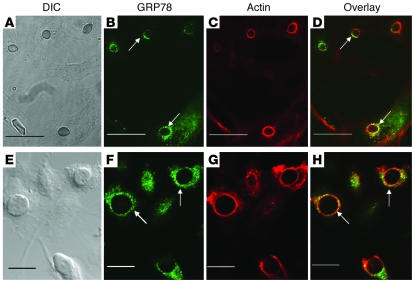
Figure 2. GRP78 on intact endothelial cells colocalizes with R. oryzae germlings that are being endocytosed.
Confocal microscopic images of endothelial cells infected with R. oryzae cells that have been germinated for 1 hour (A–D) or 2 hours (E–H). Confluent endothelial cells on a 12-mm-diameter glass coverslip were infected with 105/ml R. oryzae germlings. After 60-minute incubation at 37°C, the cells were fixed with 3% paraformaldehyde, washed, blocked, and then permeabilized (31). The cells were stained with GRP78 using rabbit anti-GRP78 polyclonal Ab (Abcam), followed by a counterstain with goat anti-rabbit IgG conjugated with Alexa Fluor 488 (Molecular Probes, Invitrogen) (B and F). To detect F-actin, the cells were incubated with Alexa Flour 568–labeled phalloidin (Molecular Probes) per the manufacturer’s instructions (C and G). A merged image is shown in D and H. (A and E) The same fields taken with differential interference contrast imaging. Arrows indicate GRP78 and microfilaments that have accumulated around R. oryzae. Scale bars: 30 μm (A–D) and 20 μm (E–H).

