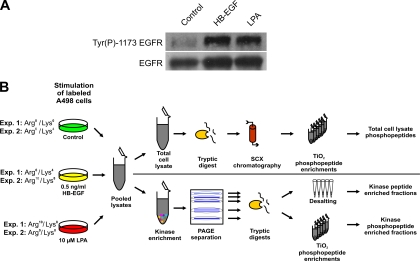
Fig. 1.
Stimulation conditions and experimental design. A, LPA- and HB-EGF-induced tyrosine phosphorylation of the EGFR in SILAC-encoded A498 cells. Immunoblot analysis of total cell extracts with antibody recognizing Tyr(P)1173 in the EGFR revealed similar stimulation upon treatment with either 0.5 ng/ml HB-EGF or 10 μm LPA. EGFR levels were similar as verified by immunoblotting with anti-EGFR antibody. B, schematic illustration of the integrated proteomics work flow for quantitative phosphorylation analysis of total lysates and kinase-enriched fractions. pY, phosphotyrosine.

