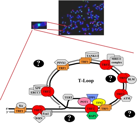
Fig. 1.
Fluorescent in situ hybridization reveals presence of telomeres at termini of human chromosomes. Top panel, representative metaphase spread from human cells. FISH analysis reveals the presence of telomeres (red) and centromeres (green), and chromosomal DNA (blue) was detected by DAPI staining. Bottom panel, schematic drawing of a telomere loop (T-Loop) showing the shelterin core complex (TRF1, TRF2, POT1, TIN2, RAP1, and TPP1) as well as a subset of known telomere-binding proteins (in gray). Question marks indicate that more telomere-binding proteins remain to be identified. WRN, Werner, BLM, Bloom, and XPF, xeroderma pigmentosum type F.

