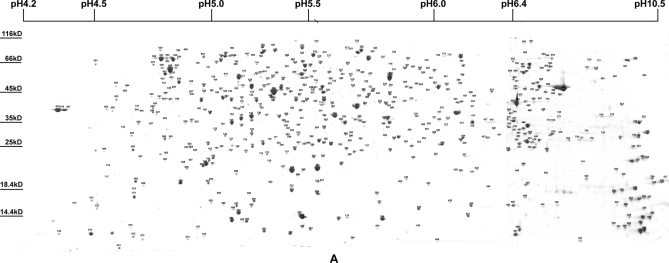
Fig. 1.
Two-dimensional gel electrophoresis and identified spots of whole-cell proteins at 37 °C in stationary phase S. flexneri. The identified spots are labeled on the integrated 2-DE map. The whole set of four maps is shown in supplemental Fig. 1 and can be found at the Proteome 2D-PAGE Database.

