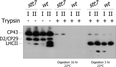
Fig. 2.
Shaving of thylakoid phosphorylated proteins using trypsin. Isolated thylakoids from wt and stt7 cells in state 1 (I) and state 2 (II) were treated with (+) or without (−) trypsin at 22 °C for 3 or 16 h for achieving partial or complete digestion, respectively. The proteins were subsequently separated by SDS-PAGE and immunoblotted with an anti-phosphothreonine antibody. The identity of the phosphorylated proteins is indicated on the left.

