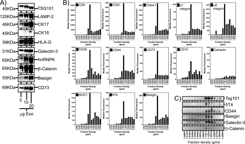
Fig. 4.
Validation of some MS-identified proteins by Western blot and flow cytometric analysis. HT1376 exosomes (5–20 μg/well), purified by the standard sucrose cushion method, were analyzed by Western blot for expression of a range of MS identified proteins as indicated (A). The 70,000 × g pellet, obtained from HT1376 cell-conditioned medium, was subjected to fractionation by centrifugation on a linear sucrose gradient (0.2–2.5 m). Fifteen total fractions were collected, and the density was measured by refractometry. Thereafter, one-third of each fraction was coupled to latex beads followed by flow cytometric analysis for exosomal surface expression as indicated (B). In parallel, the remaining two-thirds of each fraction was subjected to Western blotting for proteins as indicated (C). The data reveal proteins floating at a recognized exosomal density range (1.12–1.2 g/ml). (The data are representative of two experiments.) CK, cytokeratin.

