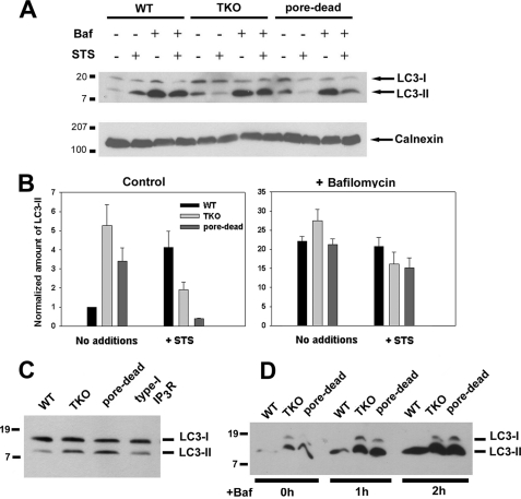
FIGURE 1.
Lack of functional IP3Rs in DT40 cells is associated with higher levels of basal autophagy. A, WT, IP3R TKO, and the D2550A pore-inactive (pore-dead) DT40 cells lines were incubated in nutrient-replete medium with serum in the presence or absence of staurosporine (1 μm) or bafilomycin A (10 nm) for 6 h. After treatment, the cells were lysed and processed for immunoblotting with LC3 Ab as described under “Materials and Methods.” The same samples were processed on 5% SDS-PAGE to monitor the levels of calnexin used as a loading control. B, levels of the LC3-II band were quantitated densitometrically and normalized to the calnexin levels. The data shown are the mean ± S.E. of 3–5 independent experiments. C, DT40 cell line stably expressing the wild-type rat type I IP3R (type I) and the functionally inactive D2550A mutant in the rat type I background (pore-dead) were directly compared for basal levels of LC3. D, WT, TKO, and pore-dead cell lines were incubated with 10 nm bafilomycin (Baf) A for the indicated times and processed for LC3 immunoblotting as described above.

