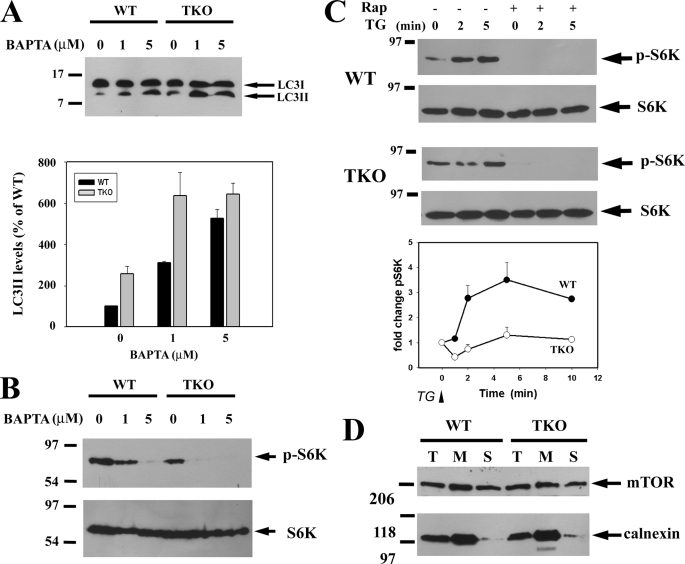
FIGURE 7.
Effect of BAPTA-AM and thapsigargin on mTORC1 activity. A, DT40 cells were incubated with the indicated concentrations of BAPTA-AM for 6 h, and the levels of LC3-II were measured by immunoblotting and quantitated in three separate experiments as shown in the bar graph. B, corresponding changes in the levels of phospho-S6 (p-S6K) kinase and total S6 kinase (S6K) from experiments carried out as in A were measured by immunoblotting. C, changes in the amount of phospho-S6 kinase was measured in DT40 cells after addition of 2 μm thapsigargin (TG). Cell lysates were prepared at the indicated times after thapsigargin treatment. When added, rapamycin (Rap, 50 nm) was preincubated with the cells for 15 min. D, DT40 cells were disrupted by five passes through a 26-gauge needle and centrifuged at 2000 × g for 5 min to remove intact cells. The total homogenate (T) was then centrifuged at 100,000 × g to obtain a membrane (M) and soluble (S) fraction. Equal protein (20 μg) from each fraction was then run on 7% SDS-PAGE and immunoblotted for mTOR. The distribution of ER protein was monitored with calnexin Ab.

