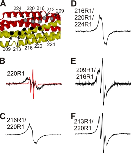
FIGURE 6.
Spin-spin interactions in R1-labeled amphiphysin dimers. A, the crystal structure of the amphiphysin dimer indicates the positions at which spin labels were introduced (Protein Data Bank code 1URU (31)). The different subunits are shown as red and gold ribbons, and the α-carbon positions of the spin-labeled sites are given as gray and black spheres. B–F, EPR spectra of indicated derivatives obtained at room temperature. The black spectra are from 100% R1-labeled proteins. The red spectrum for the 220R1 derivative was obtained at 15% R1 labeling and is shown for comparison at 10-fold reduced amplitude (B). The scan width for all spectra is 300 gauss. These spectra are normalized to the same number of spins.

