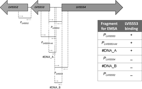
FIGURE 1.
Identification of the LVIS553 binding site. The genomic environment of LVIS553 was extracted and analyzed to identify putative binding regions. Two membrane proteins were encoded upstream and downstream of LVIS553. Different primer combinations (Table 1) were used to determine the smallest DNA binding region for LVIS553. EMSA results are summarized in the inset. +, positive binding of LVIS553 using 10 nm pure protein; −, negative binding using up to 500 nm purified LVIS553 protein.

