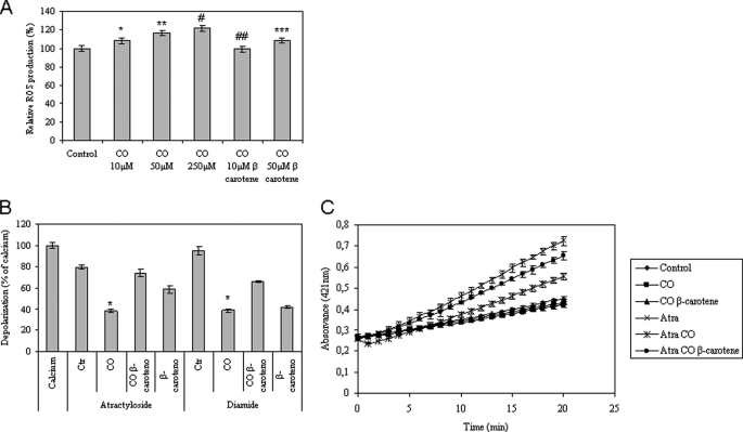
FIGURE 4.
Influence of ROS on CO effect at mitochondrial level. In A, mitochondria were treated with 10, 50, or 250 μm CO in the presence or absence of 1 μm β-carotene, followed by ROS quantification using H2DCFDA (λex, 485 nm; λem, 530 nm). The values are expressed in percentage relative to control (100%). All values are mean ± S.D., n = 4. *, p < 0.05 compared with control; **, p < 0.05 compared with control; #, p < 0.05 compared with control; ##, p < 0.05 compared with 10 μm CO; ***, p < 0.05 compared with 50 μm CO. B, mitochondria were pretreated with 1 μm β-carotene and 10 μm CO, and then atractyloside at 300 μm or diamide at 250 μm was added. The fluorescent measurements (λex, 485 nm; λem, 535 nm) are expressed in relative percentage to 5 μm Ca2+ (100%) at 15 min of incubation. All values are mean ± S.D., n = 3; *, p < 0.05 compared with control and with β-carotene and CO-treated mitochondria. C, inner membrane permeabilization was assessed according to Ref. 29. Measurements were performed at 412 nm in the absence or presence of 10 μm CO and 300 μm atractyloside for 20 min at 37 °C. All values are mean ± S.D. (error bars), n = 3.

