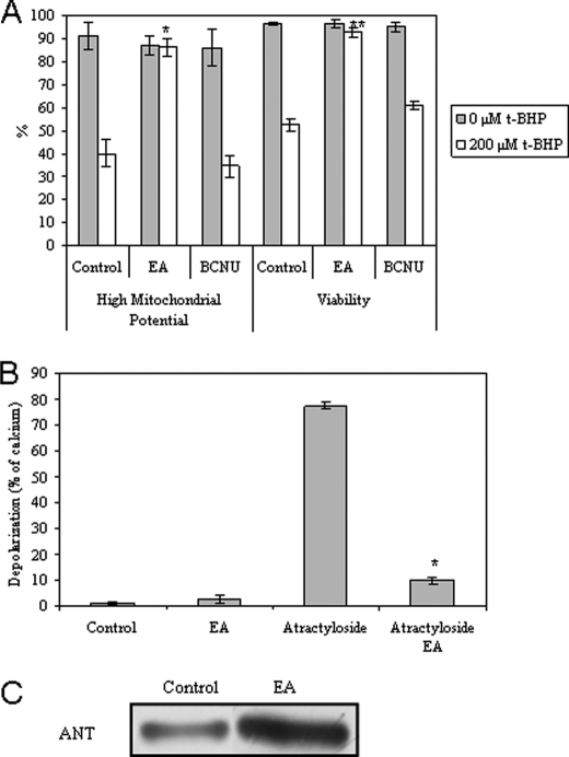
FIGURE 9.
Effect of mitochondrial GSSG on cell death, MMP, and post-translational ANT modifications. A, the percentage of astrocytic survival when subjected to 50 μm EA or 100 μm BCNU for 1 h, followed by medium exchange and treatment with t-BHP (200 μm) for 18 h. Mitochondrial potential and viability were assessed by flow cytometry, using DiOC6(3) and PI, respectively. All values are mean ± S.D. (error bars), n = 4. *, p < 0.05 compared with control cells treated with t-BHP for ΔΨm high. **, p < 0.05 compared with control cells treated with t-BHP for viability. B, depolarization assay. Isolated mitochondria were pretreated with 10 μm EA for 10 min, followed by 300 μm atractyloside or 5 μm Ca2+ treatment at 37 °C. The fluorescent measurements (λex, 485 nm; λem, 535 nm) are expressed in relative percentage to 5 μm Ca2+ (100%) at 15 min of incubation. All values are mean ± S.D., n = 4. *, p < 0.05 compared with atractyloside-treated mitochondria. C, primary cultures of astrocytes were treated with 0 or 50 μm EA following mitochondria isolation; glutathionylated proteins (α-GSH) were immunoprecipitated in mitochondria isolated from astrocytes, and ANT was immunodetected by Western blot from the immunoprecipitated proteins. This experiment was repeated three times with similar results.

