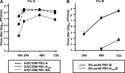
FIGURE 4.
Viral growth of FluA and FluB mutant viruses. MDCK cells were infected with the indicated FluA viruses expressing PB1-A (A/SC35M PB1-A), PB1-AT6Y (A/SC35M PB1-AT6Y), PB1-BA (A/SC35M PB1-BA) (A) or FluB viruses expressing WT PB1-B (B/Lee/40 PB1-B) or PB1-B harboring the dual PA-binding domain (B/Lee/40 PB1-AT6YB) (B) at an m.o.i. of 0.001. Error bars represent S.D. from three independent experiments. PFU, plaque-forming units; FFU, fluorescence-forming units.

