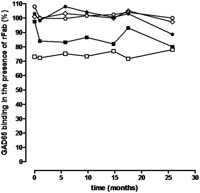
Figure 2. Epitope analysis using GAD65-specific rFab.

Longitudinal samples taken at the indicated time points were analyzed for their epitope binding pattern in our epitope-specific RBA using GAD65-specific rFab b78 (black squares), b96.11 (white squares), MICA-3 (white circles), DPD (diamonds), and N-GAD65 mAb (black circles). Binding to radiolabled GAD65 by serum samples was tested in the absence (100%) and presence of each of these rFab at half-maximal concentration.
