
Figure 3. DR301-tetramer staining of the PBMC from the SPS patient.
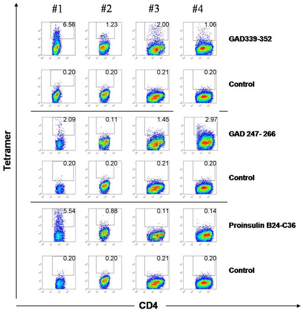
PBMC at 14-day post-stimulation with GAD65 339–352, GAD65 247–266 or proinsulin B24-C36 peptide (10 μg/ml) were stained with both specific DR301 tetramers containing the cognate peptide (upper panels in each section), and DR301-NS1 negative control tetramer (lower panel in each section). The cells were gated on live lymphocytic population and CD3+ cells. The frequency of CD4 and tetramer-positive cells is shown in the quadrant. The tetramer-positive cells were single cell sorted on 96-well plates.