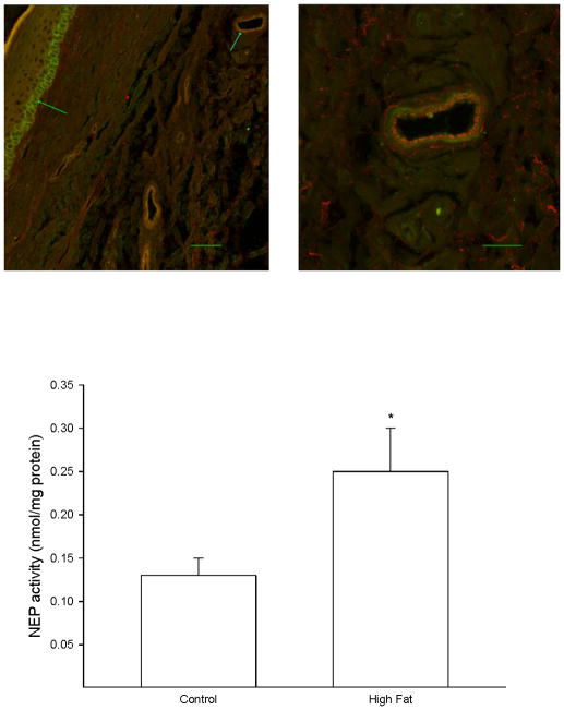
Figure 4. Effect of a high fat diet on neutral endopeptidase activity in the skin of the hindpaw.
Rats were fed a standard or high fat diet for 32 weeks. Afterwards, activity of neutral endopeptidase in a skin biopsy of the hindpaw was determined as described in the Methods section. A representative image for expression of neutral endopeptidase in the hindpaw is provided (top). The left image (inserted bar 50 μm) demonstrates the staining for neutral endopeptidase (green) that occurs in basal keratinocytes in the stratum basale (green arrow on left). The blue arrow pointing at the vessel in the upper right portion of the image on the left side is enlarged and shown on the right side. The right image (inserted bar 20 μm) demonstrates that staining for VWF occurring in the endothelium (red staining) does not overlap with the staining for neutral endopeptidase (green staining) suggesting that neutral endopeptidase in the vasculature is primarily located in the smooth muscle layer. The number of rats in each group was the same as shown in Table 1. * p < 0.05, compared to rats fed the standard diet (control).

