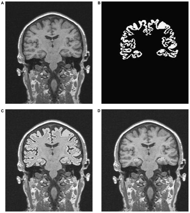
Fig. 1.

A pictorial representation of the steps involved in the hippocampus quantification routine. (1a) Sagittal MRI series is resliced into 1-mm coronal images. (1b) An example of cortical gray matter segmentation. (1c) Cortical mask “overlay” on T1 image. (1d) Hippocampus tissue extracted by trained rater utilizing manual editing to separate the body of the hippocampus from the cortex.
