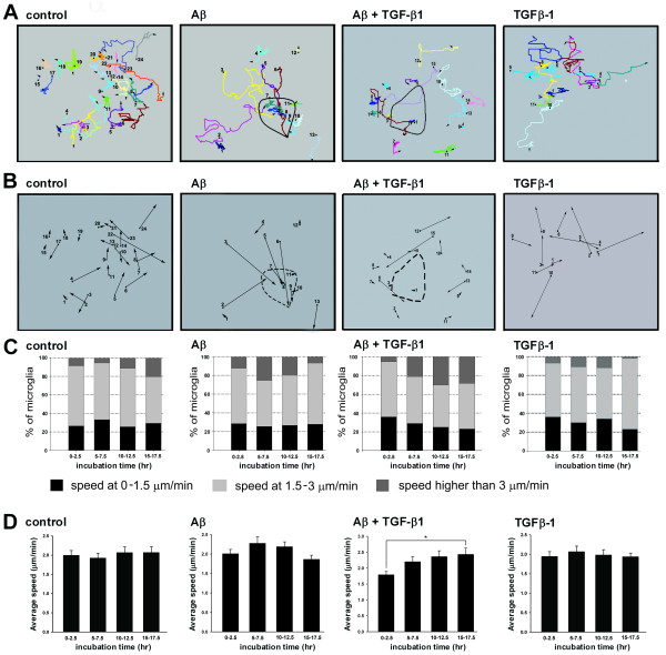
Figure 4.
Cell tracings and speed analysis of Aβ-treated microglia in the absence and presence of TGF-β1. The migratory tracks of control, TGF-β1-, and Aβ-treated BV-2 cells, with and without TGF-β1 were labeled in different colors (A). The migratory direction of BV-2 cells are represented by the vectors linking the start and end points (B). Speed distributions were analyzed during 0-2.5, 5-7.5, 10-12.5, and 15-17.5 h after the addition of Aβ aggregates (C). Black bars represent the percentage of cells with a speed between 0-1.5 μm/min. Light gray bars represent the percentage of cells with a speed between 1.5-3 μm/min. Dark gray bars represent the percentage of cells with a speed greater than 3 μm/min. Average speeds were calculated by averaging the migration speed during the entire recording period (D). Bars represent the mean ± SEM. Comparisons marked with asterisks are significant different. At least 10 cells from each independent time-lapse recording were pooled for speed distribution and average speed analysis. * = p < 0.05.

