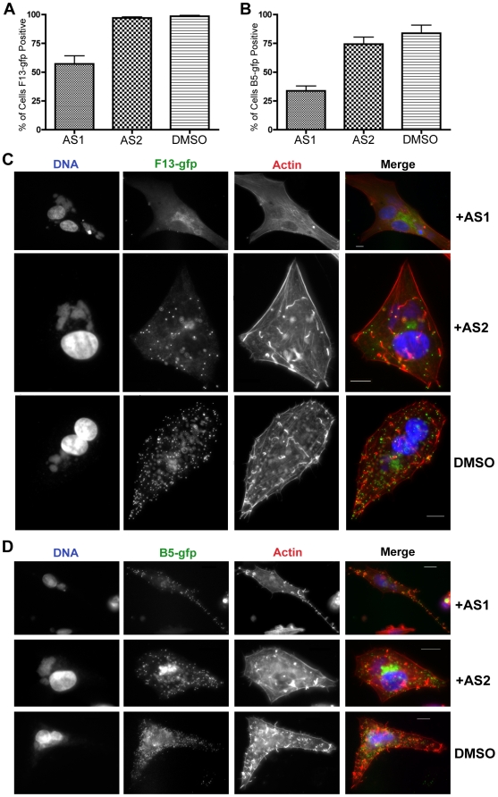
Figure 4. PI3K inhibitors alter the distribution of F13 and B5 in infected cells.
A. BHK cells were infected with VV F13-GFP, and treated with AS1 (50 µM) or AS2 (50 µM) post-adsoption for 16 hours. Cells were fixed and stained with phalloidin to visualize actin (red) and DAPI to visualize DNA (blue). Approximately 600 cells were counted per condition and cells were scored positive if a FITC (GFP) signal could be detected. B. BHK cells were infected with VV B5-GFP, and treated with PI3K inhibitors post adsoption for 16 hours, and stained as in A. Approximately 400 cells were counted per condition. C. PI3K inhibitors alter the distribution of F13-GFP in infected cells. Cells were infected and stained as in A. Note that AS1 and AS2 cause a reduction of punctate FITC fluorescence at the cell periphery, which represents virions, as well as a reduction in the number of actin tails. AS1 also caused F13-GFP to localize to the cytoplasm instead of on punctate virions. D. PI3K inhibitors alter the distribution of B5-GFP in infected cells. Cells were infected and stained as in B. Note that AS1 and AS2 had similar effects on localization of B5 and F13, namely a reduction in peripheral punctate virions and reduced numbers of actin tails. Results are from three independent trials.

