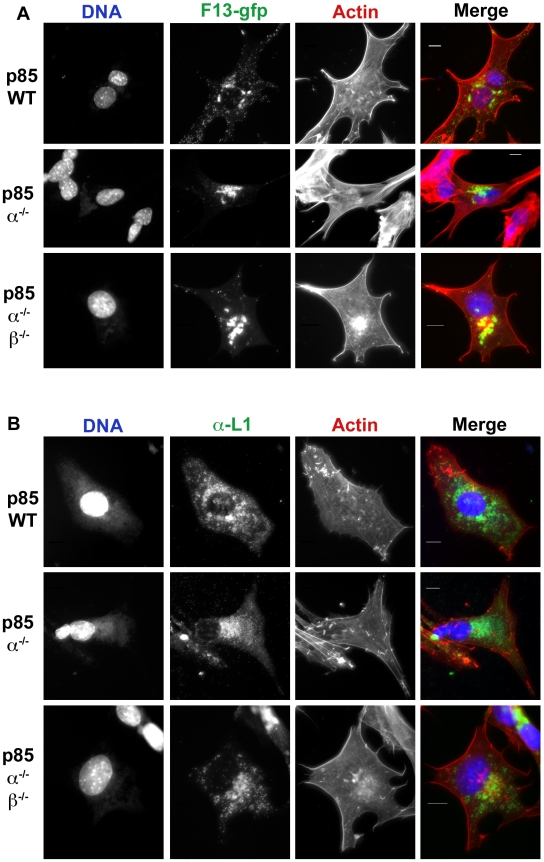
Figure 6. Localization of F13-GFP and L1 in p85-deficient cells.
A. p85-deficient cells were infected with VV F13-GFP for 16 hours. Cells were fixed and stained with phalloidin to visualize actin (red) and DAPI to visualize DNA (blue). Note that in the p85α−/− and p85α−/−β−/− cells there is a reduction of punctate FITC fluorescence at the cell periphery, which represents virions, as well as a reduction in the number of actin tails. F13 remains in a peri-nuclear location in p85-deficient cells. B. L1 production and localization in p85-deficient cells. p85-deficient cells were infected with VV, strain WR for 16 hours. Cells were fixed and stained with phalloidin to visualize actin (red), DAPI to visualize DNA (blue), and L1 antibody, followed by FITC conjugated anti-Mouse antibody. No difference in L1 localization the p85-deficient cells compared to wild type cells was apparent. Results are from three independent trials.

