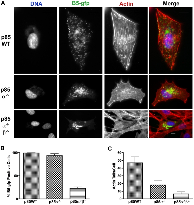
Figure 7. Localization of B5-GFP in p85-deficient cells.
A. p85-deficient cells were infected with VV B5-GFP for 16 hours. Cells were fixed and stained with phalloidin to visualize actin (red) and DAPI to visualize DNA (blue). Note that in the p85α−/− and p85α−/−β−/− cells there is a reduction of punctate FITC fluorescence at the cell periphery, which represents virions, as well as a reduction in the number of actin tails. B5 remains in a peri-nuclear location in p85-deficient cells. B. p85-deficient cells were infected as in A and percent B5-GFP cells were quantified. Approximately 400 cells were counted per condition and cells were scored positive if a FITC (GFP) signal could be detected. p85α−/−β−/− cells had significantly reduced rates of B5-GFP positive cells. Results are from three independent trials. C. p85-deficient cells form fewer actin tails than p85WT cells. Actin tails were counted for ∼10 cells per condition. Data is from one representative experiment.

