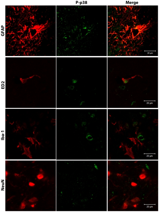
Figure 9. Phospho-p38 is expressed in astrocytes and perivascular microglia.
Confocal analysis was used to determine phospho-p38 (P-p38) cellular localization in the superficial laminae of the ipsilateral L5 dorsal horn in AM281 + AM630-treated rats on day 15 after paw incision. Representative images are shown. Phospho-p38 appears in green. GFAP (marker for astrocytes), ED2/CD163 (ED2, marker for perivascular microglia), Iba-1 (marker for microglia) and NeuN (marker for neurons) appear in red. The color of ED2/CD163, Iba-1 and NeuN staining was digitally changed from green to red, and phospho-p38 from red to green for consistency in presenting the data and in order to allow a better visualization of the co-localization of these markers. The colors of the GFAP/phospho-p38 co-stain (top panel) were not altered.

