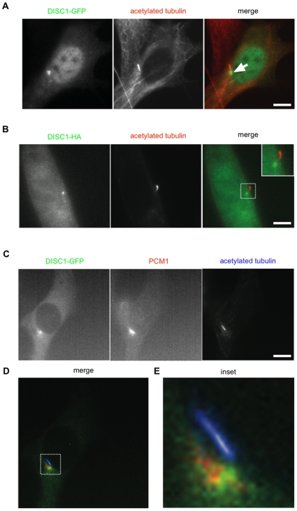
Figure 1. Localization of DISC1-GFP near the base of primary cilia.
(A) NIH3T3 cells transfected with DISC1-GFP were fixed and immunolabeled for acetylated tubulin to mark primary cilia. An example of such a double-labeled cell is shown, with DISC1-GFP in green and acetylated tubulin in red in the merged image. Arrow indicates indicates the concentration of DISC1-GFP observed near the ciliary base. (B) The same experiment conducted using DISC1-HA. (C) Triple localization of DISC1-GFP, endogenous PCM1, and acetylated tubulin verifying localization of DISC1 in a centrosomal region near the ciliary base. (D) Merged image from the triple localization with DISC1-GFP in green, PCM1 in red, and acetylated tubulin in blue. (E) The region indicated in panel D displayed at higher magnification. Scale bar, 10 µm.

