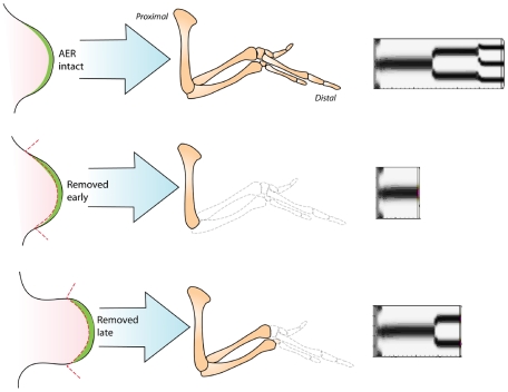
Figure 4. Simulations of AER removal.
(Left two columns) Drawings of AER removal experiments, based on Saunders's study [40]. Top images show an intact chicken wing bud at an early stage of development and the limb skeleton that it generates. Middle images show a wing bud at the same early stage with the AER removed, and the resulting limb skeleton, which attains a normal size but is truncated beginning at the elbow. Bottom images show a later stage wing bud whose AER has been removed. The resulting skeleton is truncated from the wrist onward. (Right column) Simulations of limb development using standard parameters. Top: AER (i.e., the source of suppressive FGF morphogen) left intact; normal development results. Middle: AER deleted early during the simulation. Bottom: AER deleted later during the simulation. All simulated limbs were allowed to develop for the same time.

