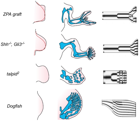
Figure 5. Simulations of effect of limb bud distal expansion.
ZPA graft (left) expanded chicken wing bud that results from anterior graft of an ectopic zone of polarizing activity from the proximal posterior region of another wing bud (normal limb profile at this stage shown in red); (center) resulting cartilage skeleton, with mirror-image duplication; (right) end-stage of simulation with distal expansion corresponding to that shown on left. Shh−/−, Gli3−/− (left) expanded mouse forelimb bud in embryos null for both Shh and Gli3 (normal limb profile at this stage shown in red); (center) resulting skeleton, with supernumerary digits [61]; (right) end-stage of simulation with distal expansion corresponding to that shown on left. talpid 2 (left) expanded wing bud of chicken embryo homozygous for talpid 2 mutation; (center) cartilage skeleton formed from such a limb bud later during development; (right) end-stage of simulation with distal expansion corresponding to that shown on left. Dogfish (left) shape of the pectoral fin-bud in an embryo of the dogfish Scyliorhinus torazame; (center) cartilaginous fin skeleton formed from such a limb bud [5]; (right) end-stage of simulation using a limb bud contour like that shown on left. In each of these simulations reaction parameter values different from the standard ones were used (see File S4 for details).

