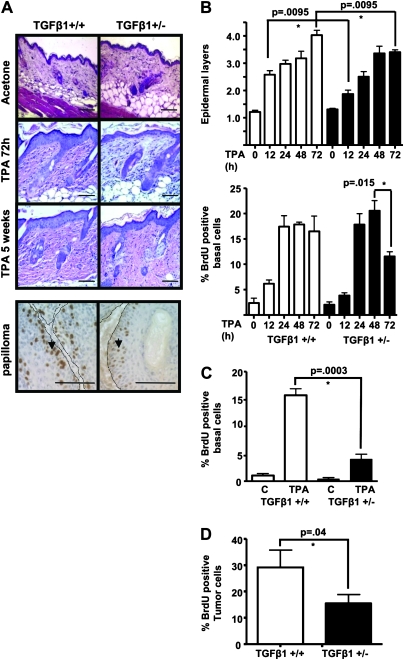
Fig. 2.
TGFβ1 enhances TPA-induced proliferation in normal epidermis and tumors. (A) Top: representative hematoxylin-and eosin-stained sections of acetone or TPA-treated skin at 72 h post treatment and after 5 weeks chronic TPA treatment, magnification ×200. Bottom: detection of proliferating tumor cells (arrows) with anti-bromodeoxyuridine (BrdU) immunohistochemistry in TGFβ1+/+ and TGFβ1+/− 10 week papillomas. Magnification ×400. Tumor basement membrane indicated by dashed line. Scale bars represent 20 μm for all images. (B) Quantitation of epidermal hyperplasia (top) and proliferation (bottom) in TPA-treated TGFβ1+/+ mice compared with TGFβ1+/− mice. The number of cell layers in hematoxylin- and eosin-stained sections was determined every 20 basal cells along a section and averaged from 5 mice per group. BrdU-positive cells were quantitated from anti-BrdU-stained sections and averaged from 5 mice per time point for each genotype. (C) Quantitation of epidermal proliferation in TGFβ1+/+ and TGFβ1+/− skin after chronic TPA treatment. BrdU-positive epidermal keratinocytes were quantitated from mice treated twice per week with TPA or acetone (c) for 5 weeks (N = 5 mice per group). (D) Quantitation of tumor cell proliferation. BrdU-positive tumor cells were quantitated from papillomas generated in each genotype with DMBA and 10 weeks TPA promotion (N = 5 tumors per group). Papillomas were isolated 72 h after last TPA treatment. Asterisk represents significantly different from indicated group.

