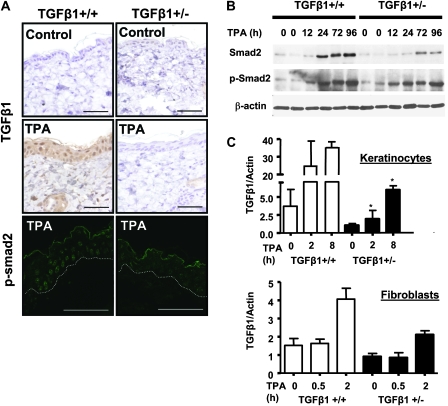
Fig. 3.
TPA induction of TGFβ1 and p-Smad2 nuclear localization is reduced in TGFβ1+/− epidermis. (A) Top and middle: immunohistochemical detection of TGFβ1 protein in skin 12 h after acetone or TPA treatment, (magnification ×400). Bottom: detection of phospho-Smad2 by indirect immunofluorescence with an anti-pSmad2 ser 465/467 antibody in tissue 24 h after TPA treatment. Magnification ×1000 and scale bars represent 20 μm. Exposure times for TGFβ1+/+ and TGFβ1+/− skins were identical. Figure S1A available at Carcinogenesis Online shows individual and merged images with TO-PRO 3 nuclear counterstaining. Location of the basement membrane is indicated by a dashed line. (B) Immunodetection of total Smad2 and p-Smad2 in whole skin protein extracts isolated at the indicated time points after TPA treatment using specific anti-Smad2/3 and p-Smad2 antibodies and β-actin as a loading control. (C) Quantitative reverse transcription–polymerase chain reaction analysis of TGFβ1 mRNA induction by TPA (25 ng/ml) in primary keratinocytes (top) and fibroblasts (bottom) of the indicated genotype. Results are the average of three independent experiments. Asterisk represents significantly different from TGFβ1+/+ (P < 0.05).

