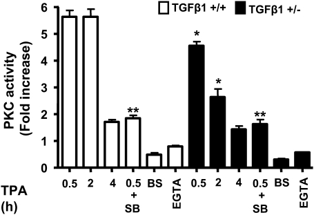
Fig. 4.
TGFβ1 modulates PKC activation by TPA. PKC enzyme activity was measured at the indicated times after TPA treatment (25 ng/ml) in triplicate cultures of primary keratinocytes of each genotype and expressed as fold increase over untreated control (1751.5 ± 256 c.p.m. for TGFβ+/+ and 1642.2 ± 171.19 c.p.m. for TGFβ+/− cells). TGFβ1+/+ keratinocytes were also pretreated for 15 min with the small molecule ALK5 inhibitor SB431542 (0.5 μM) before addition of TPA, and PKC activity was measured after 30 min. Similar results were obtained in three independent experiments. Bisindolylmaleimide I (BS) and ethyleneglycol-bis(aminoethylether)-tetraacetic acid (EGTA) were included in the activity assay to inhibit total and calcium-dependent PKC activity, respectively. Asterisk represents significantly different from indicated group.

