
Figure 1.
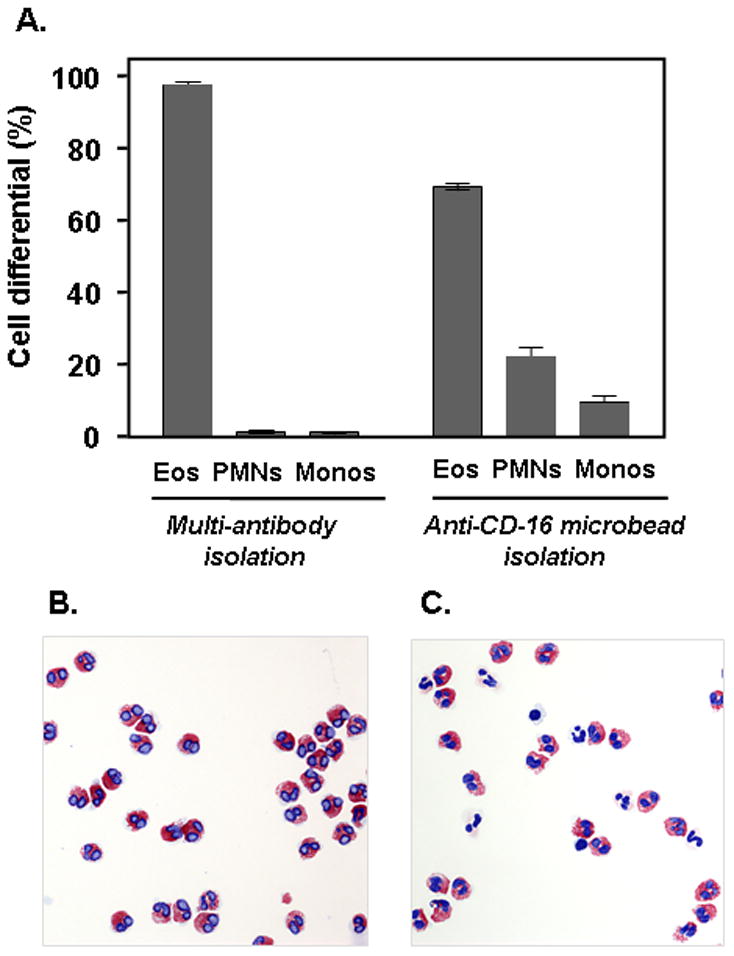
(A) Leukocyte differentials after isolation of eosinophils using the Milltenyi multi-antibody method compared to that obtained after isolation using the original anti-CD16 microbeads alone. Eos, eosinophils; PMNs, polymorphonuclear leukocytes, or neutrophils; Monos, monocytes. Differentials shown are based on 500 leukocytes counted for each of two separate isolations for each method; representative experiment of n = 3. (B) and (C) are cytospin preparations of cells from typical multi-antibody and anti-CD16 microbead isolations, respectively (modified Giemsa stain, original magnification, 20X).