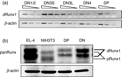
Figure 1.
Expression profiles of Runx1 transcript and protein in the double-negative (DN) and double-positive (DP) fractions prepared from thymocytes of wild-type C57BL/6 mice. (a) Semi-quantitative reverse transcription–polymerase chain reaction (RT-PCR) analysis of distal Runx1 transcript levels during thymocyte differentiation. RNA was extracted from DN1/2, DN3S, DN3L, DN4 and DP fractions each, and converted to complementary DNAs (cDNA). An increasing amount of cDNA was used for PCR as indicated. Transcript of β-actin served as a control. (b) Immunoblot analysis of Runx1 protein in thymocytes. Protein extracts were prepared from the DN and DP fractions as well as from EL-4 and NIH3T3 cell lines, and processed for immunoblot detection using anti-panRunx antibody. A slight but significant difference in the migration corresponded to distal and proximal Runx1 polypeptides as indicated. β-actin served as a loading control.

