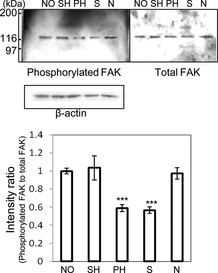
FIGURE 2.

Amounts of phosphorylated and total FAK in rHSCs. rHSCs were detached and replated on dishes coated with NO-, SH-, PH-, desialylated, or de-N-glycosylated VN (10 μg/ml) for 90 min. The cell lysates were immunoblotted with anti-phosphotyrosine antibody (PY-20) to detect phosphorylated FAK as described in the text. Immunostaining of phosphorylated FAK, total FAK, and β-actin is shown in the upper panels. The relative amount of phosphorylated FAK, normalized to total FAK, was calculated by using the software program Scion Image and expressed by taking the intensity of the band of NO-VN as 1. The data represent the means ± S.D. (n = 3, meaning three separate experiments) and were analyzed by Student's t test; ***, p < 0.001 compared with NO-VN. Abbreviations: S, desialylated VN; N, de-N-glycosylated VN.
