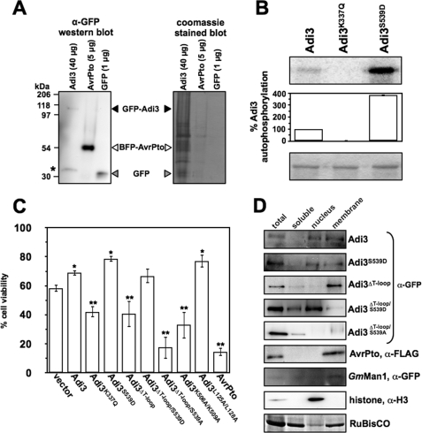
FIGURE 2.
Cellular localization of GFP-Adi3 proteins regulates cell death suppression (CDS) activity. A, GFP-Adi3 protein expression levels under the 35S promoter. Tomato protoplasts were transformed with the indicated DNA constructs and protein expressed for 16 h followed by total protein extraction and α-GFP Western blot analysis. B, GFP-Adi3 autophosphorylation activity. Recombinant GFP-Adi3 protein was immunoprecipitated using α-GFP-agarose and tested in an in vitro kinase assay as in Fig. 1B. Values are reported as a percentage of wild-type Adi3 autophosphorylation and are average of three independent experiments. Error bars are standard error. Top panel: kinase assay (phosphorimage). Bottom panel: assay input (Coomassie Blue-stained gel). C, cell viability of protoplasts expressing GFP-Adi3 plus NLS and NES mutants. Tomato protoplasts expressing the indicated proteins for 24 h were analyzed for cell viability by Evans Blue staining to identify dead cells. Data represent the average of five independent experiments. Error bars are standard error. One asterisk and two asterisks indicate significant increase or decrease, respectively, in cell viability compared with vector-only expression (Student's t test, p < 0.05). D, subcellular distribution of GFP-Adi3 proteins. The indicated proteins were expressed for 16 h in tomato protoplasts followed by subcellular fractionation and Western blot with the indicated antibodies. Markers used: membrane, AvrPto-FLAG (73) and soy bean α-1,2-mannosidase (GmMan1)-mCherry (35); nuclei, histone H3; soluble fraction and loading control, RuBisCo (Coomassie Blue stain of membrane). Western blots are representative of three independent experiments.

