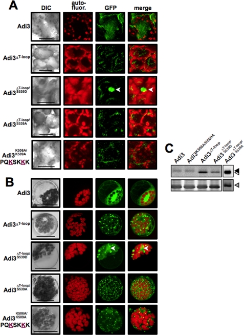
FIGURE 5.
Adi3 NLS mutants are located in punctate cellular structures. A, localization of GFP-Adi3 proteins in intact PtoR tomato leaf mesophyll cells. After Agrobacterium-mediated transient expression for 48 h (GFP-Adi3), 24 h (GFP-Adi3ΔT-loop; GFP-Adi3ΔT-loop/S539D), or 12 h (GFP-Adi3ΔT-loop/S539A; GFP-Adi3K506A/K509A), leaf sections were cut from 3 different leaves and visualized by confocal microscopy. Images are representative of three independent experiments. Merge, overlay of GFP and autofluorescence images. Bar, 20 μm; arrowhead, nucleus. B, localization of GFP-Adi3 proteins in PtoR tomato protoplasts. The indicated GFP-Adi3 proteins were expressed in protoplasts for 16 h and visualized by confocal microscopy. Labeling as in A. C, Western blot using α-GFP antibody on total protein from expression of the indicated GFP-Adi3 proteins. Filled triangle, GFP-Adi3; open triangle, GFP-Adi3ΔT-loop proteins; gray triangle, RuBisCo loading control (Coomassie Blue-stained membrane).

