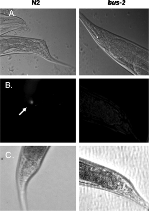
FIGURE 1.
Microscopy images of N2 Bristol and bus-2 strains cultured with M. nematophilum. The ventral view is shown. A, shown is Nomarski differential interference contrast of C. elegans grown on a mixed culture of M. nematophilum and E. coli. The dar phenotype is present in wild type but not bus-2 worms. B, SYTO green stained bacteria are seen in the anal region of N2 Bristol but not bus-2 nematodes; the arrow points to localization of florescence in the anal region. C, the light microscopy counterpart of fluorescence images seen in B is shown.

