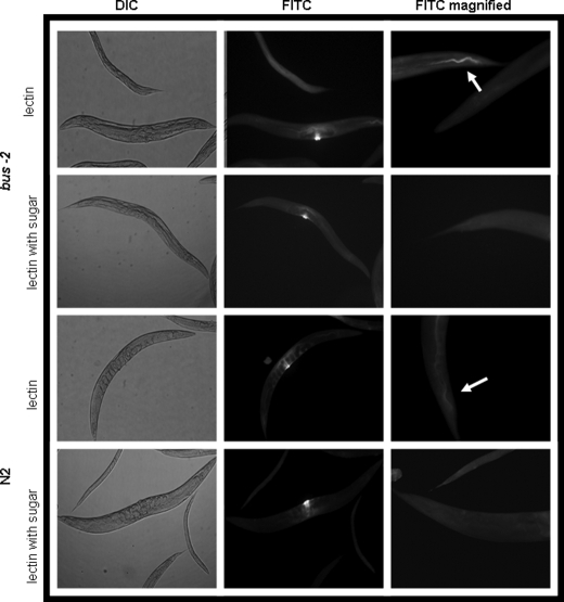
FIGURE 5.
UEA I α-l- fucose specific lectin staining of whole mounted delipidated adult C. elegans. The left column shows control differential interference contrast (DIC) micrographs of lectin-treated nematodes with and without prior incubation with inhibitory concentrations (1 mm) of α-l-Fuc. The images in the center column show that vulva staining is background, as it is not affected by preincubation with α-l-Fuc. The right column shows fluorescence micrographs of UEA I. The bus-2 strains contain a higher abundance of fucosyl glycoproteins. FITC, fluorescein isothiocyanate.

