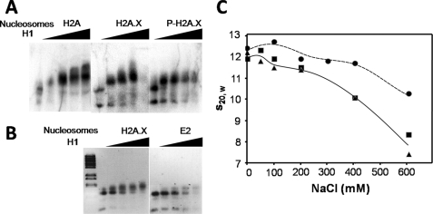
FIGURE 6.
Impaired binding of linker histones to nucleosomes by H2A.X and γ-H2A.X. A, nucleosomes reconstituted with H2A, H2A.X, and P-H2A.X and [γ-32P]ATP-labeled 208-bp DNA were titrated with increasing amounts of histone H1 and analyzed by 1% agarose gel electrophoresis. The histone H1 to nucleosome molar ratios used were 0, 0.5, 1.0, 1.5, and 2.0. The image shows an autoradiograph of the gel. B, shown is an ethidium bromide-stained image of a similar H1-dependent gel shift carried out in a 1% agarose gel using 208-bp reconstituted nucleosomes consisting of H2A.X and H2A.X-A138E/S139E. C, shown is sodium chloride concentration dependence of the sedimentation coefficient (s20,w) of 208-bp reconstituted nucleosomes consisting of native H2A (black circles), H2A.X (black squares), and P-H2A.X (black triangles).

