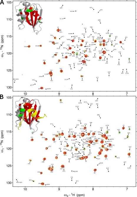
FIGURE 2.
Deuterium solvent-exchange protection in VAMP7(1–118) and VAMP7(1–180). A, overlay of 1H, 15N-HSQC spectra of VAMP7(1–118) after 1 min and 25 s (green), 6 min and 15 s (orange), and 11 and 5 min and 5 s (red) from resuspension in deuterated solvent. Top left corner, the detectable resonance peaks of these three spectra are mapped on the schematic of the LD-Hrb136–176 crystal structure (PDB ID 2VX8) (32) using the colors corresponding to the three spectra. The LD peaks that were not detected due to rapid deuterium exchange are colored gray. B, overlay of 1H, 15N-HSQC spectra of VAMP7(1–180) after 1 min and 20 s (green), 6 min and 5 s (orange), and 11 min (red) from resuspension in deuterated solvent. Top left corner, the detectable resonance peaks of these three spectra are colored in the LD-Hrb136–176 as described for panel A.

