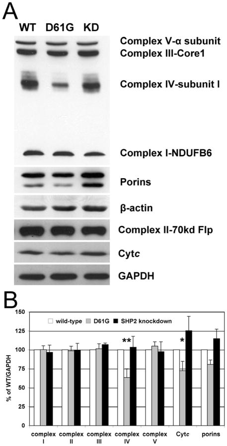
Fig. 2.
Expression analysis of OxPhos complexes and cytochrome c. (A) Western blot analysis of primary antibodies were as follows. Complex I: anti-NDUFB6, complex II: anti-70kd flavoprotein, complex III: anti-core I, complex IV: anti-subunit I, complex V: anti-α subunit, GAPDH: glyceraldehyde 3-phosphate dehydrogenase. (B) Quantitative assessment of protein levels by densitometric analysis of Western blots normalized to GAPDH (n = 3). Among OxPhos components, CcO and Cytc levels are 37% (p<0.02, **) and 28% (p=0.05, *) reduced in D61G cells.

