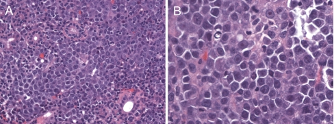
Fig. 2.
Hematoxylin and eosin stained sections. a (original magnification ×200) There is a monotonous population of malignant cells diffusely infiltrating salivary gland parenchyma. b (original magnification ×400) The malignant cells have a plasmablastic appearance with clumped chromatin, often a single prominent “cherry red” nucleolus, smooth nuclear contours, and eccentric cytoplasm

