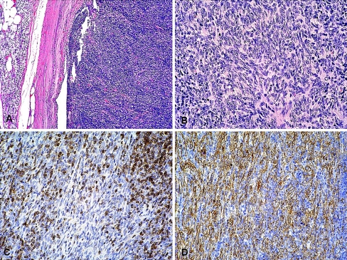
Fig. 3.
Case 1. Photomicrographs (Hematoxylin and Eosin-stained sections). a Encapsulated tumor with a predominant lymphocyte-rich component. A rim of normal parathyroid tissue is present external to the capsule on the left (×100). b Lymphocyte-poor spindle cell area. The spindle cells show minimal cytologic atypia (×200). The Immunohistochemical study shows c positive reaction for CD1a in non-neoplastic lymphocytes and d positive reaction for cytokeratins (MNF116) in neoplastic cells (×200)

