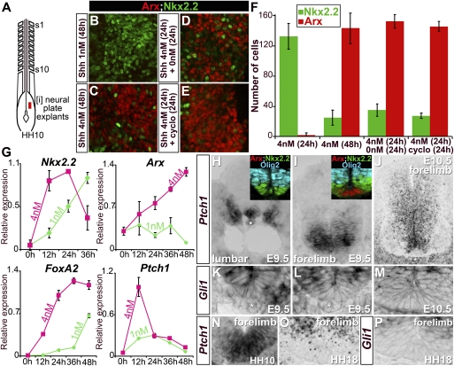
Figure 2.
Induction of FP markers by transient high levels of Shh signaling. (A) Schematic of HH10 chick embryos indicating the position of [i] neural plate explants. (B–E) Expression of Arx and Nkx2.2 in [i] neural plate explants after 48 h of culture. Cells in explants treated with 1 nM Shh exclusively express Nkx2.2 (B), while 4 nM Shh promotes FP fate, identified by Arx expression (C). In explants first exposed for 24 h to 4 nM Shh and then placed in a media either devoid of Shh (D) or containing 500 nM cyclopamine (cyclo) (E), FP induction is retained. (F) Quantification of cells expressing Arx and Nkx2.2 in [i] neural plate explants after 24 or 48 h of culture (n ≥ 4 explants, number of cells per unit ± SD). (G) Temporal dynamics of Nkx2.2, Arx, FoxA2, and Ptch1 expression relative to Actin transcripts and normalized between sets of experiments assayed by quantitative PCR in [i] neural plate explants treated with 1 nM (pale green) or 4 nM (pink) Shh (n ≥ 2 experiments, mean ± SD). As definitive FP markers are induced, p3 markers and Ptch1 are down-regulated. (H–P) Expression of Ptch1 (H–J,N–O) and Gli1 (K–M,P) in mouse embryos (H–M) and chick embryos (N–P). Insets in H and I indicate Arx, Nkx2.2, and Olig2 expression on sections adjacent to the main panel. Midline cells transiently express high levels of Ptch1 and Gli1. The asterisks indicate the notochord.

