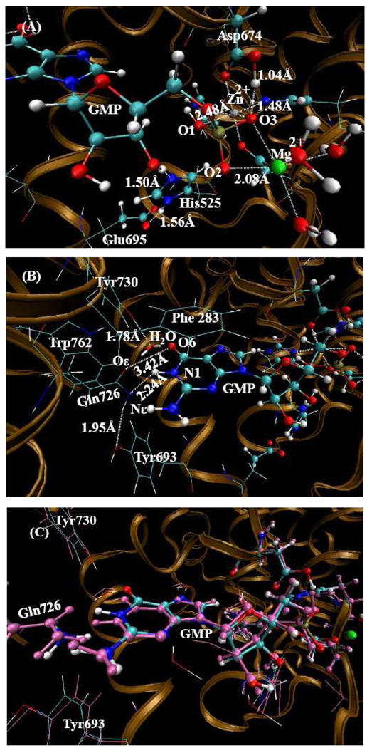
Figure 3.
QM/MM-optimized PDE10-GMP structure (the charge on GMP is −1) at the B3LYP/6-31G*:Amber level. The QM atoms in the PDE10 active site are represented by balls. All MM atoms are represented as lines. (A) From this orientation, one can see the metal sites and the state of GMP. (B) From this orientation, one can see the hydrogen bonding network around Gln726 residue and one weak hydrogen bond between Gln726 and guanine group of GMP. (C) The comparison between the QM/MM-optimized structure and the X-ray crystal structure while maintaining the original amide position of Gln726. The pink atoms represent the X-ray crystal structure added H-atoms, and the remaining atom-types represent the QM/MM-optimized structure.

