Figure 2.

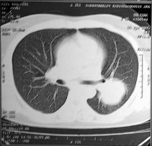
Computed tomography (CT) scan shows a well defined mass in the left lung; there was no hilar or mediastinal lymphadenopathy in the mediastinal windows

Computed tomography (CT) scan shows a well defined mass in the left lung; there was no hilar or mediastinal lymphadenopathy in the mediastinal windows