
Figure 3. UVRAG recruits C-Vps protein to the GFP–LC3-labelled autophagosomes.
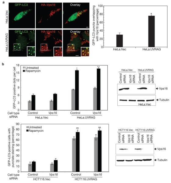
(a) HeLa.Vec and HeLa.UVRAG cells transfected with HA–Vps16 and GFP–LC3 were treated with 2 μM rapamycin for 2 h, and processed for confocal microscopy (left panel; scale bars, 5 μm). The percentage of GFP–LC3 punctae positive for HA–Vps16 staining was quantified (right panel; data are mean ± s.e.m., n = 60, *P < 0.01). (b) Effects of Vps16 on UVRAG-mediated autophagosome formation. Light microscopic quantification of autophagosomes in HeLa.Vec and HeLa.UVRAG cells (upper left panel) or in HCT116.Vec and HCT116.UVRAG cells (bottom left panel) when transfected with GFP–LC3 together with control siRNA or Vps16 siRNA (data are mean ± s.e.m., n = 60 for HeLa cells; n = 450 for HCT116 cells, *P < 0.01; **P < 0.001). Immunoblotting of Vps16 and tubulin are shown in the right panel.